
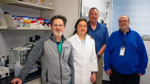
The University of Iowa Department of Ophthalmology has long been a leader in advancing vision science, and today, its cornea division is setting new benchmarks in translational and clinical research. Dr. Mark Greiner, a clinician-scientist and corneal specialist at Iowa, is at the forefront of a wide-ranging research portfolio aimed at tackling major challenges in corneal disease—particularly Fuchs endothelial corneal dystrophy (FECD), corneal transplantation in diabetic patients, and parasitic infections like Acanthamoeba keratitis.
“We’re positioning ourselves to not just participate in research but lead it—both at the bench and at the bedside,” says Dr. Greiner. A central focus is Fuchs dystrophy, the most common indication for corneal transplantation. While current treatment is limited to surgical intervention, Dr. Greiner and his colleagues are laying the groundwork for a future in which medical therapies may prevent or delay the need for surgery altogether.
One major initiative is Iowa’s involvement in the DTX natural history trial, which is investigating the progression of Fuchs dystrophy. Alongside this clinical collaboration, Dr. Greiner’s lab is leading NIH-funded work on rho kinase (ROCK) inhibitors, a class of drugs more commonly associated with glaucoma treatment. His team is conducting a head-to-head comparison of netarsudil and ripasudil—studying not only how these drugs accelerate healing after Descemet stripping but also whether they can stabilize the cornea without surgery. “It’s still the Wild West with these medications; we don’t yet know the optimal dosing, side effect profile, or long-term impact,” he notes. However, the potential to shift from surgical to pharmacological management is driving intense interest.
The lab’s second NIH grant focuses on ferroptosis, a recently recognized mechanism of cell death implicated in Fuchs dystrophy. By targeting this pathway with ubiquinol (the active form of Coenzyme Q10), the team hopes to prevent or slow cellular degeneration in the corneal endothelium. “We’re building the pipeline for future human trials,” Greiner explains. “If these molecules continue to perform well in preclinical models, we’ll be pushing toward FDA approval in the years ahead.”
Complementing this molecular work are projects aimed at understanding the real-world impact of Fuchs dystrophy. Dr. Chris Salas, in collaboration with optometrist Dr. Mark Wilkinson, conducted a study using the National Advanced Driving Simulator to measure how Fuchs affects functional vision, even in patients who are 20/20 on acuity charts. The study found that patients with Fuchs—despite excellent visual acuity—reported feeling unsafe while driving and demonstrated quantifiable impairments behind the wheel. These findings underscore the need for early intervention and give clinical relevance to Iowa’s therapeutic development work. “We now have a bridge between molecular changes and patient-reported outcomes,” says Greiner.
In another forward-thinking project, the team is developing software to measure and track guttae accumulation in specular microscopy images. This technology could allow clinicians and researchers to more precisely monitor disease progression and treatment response. “It’s the corneal equivalent of drusen in macular degeneration,” Greiner explains. The tool is currently in the process of being licensed to major specular microscope manufacturers, with the goal of making it widely available to investigators and clinicians.
Outside of Fuchs dystrophy, Dr. Greiner’s lab has been deeply involved in understanding how diabetes affects corneal transplant outcomes. What began as an observation by Iowa Lions Eye Bank’s Greg Schmidt—who noticed DMEK grafts from diabetic donors were more likely to tear—has evolved into a body of published research and a multicenter clinical trial: the Diabetes Endothelial Keratoplasty (DEX) Study. This randomized, prospective trial examines graft success, endothelial cell survival, and morphological changes over time. Iowa researchers played a key role from the initial observation through to study design and executive committee leadership. The first results are expected to be published around the upcoming AAO meeting and may have major implications for donor tissue selection and surgical outcomes worldwide.
Iowa’s leadership continues with the Parasitic Ulcer Treatment Trial (PUTT), another multicenter randomized clinical trial, this time investigating how topical steroids influence outcomes in Acanthamoeba keratitis. Steroids are often used empirically in presumed infectious keratitis, but their effect in Acanthamoeba infections—rare but potentially blinding—is unclear. Dr. Greiner explains, “By the time patients reach us, many have already been started on steroids, and we don’t know if that’s helping or hurting once definitive anti-Acanthamoeba therapy is initiated.” The PUTT study will randomize patients already receiving anti-parasitic treatment to either placebo or topical steroid, addressing this long-standing clinical question. Iowa’s leadership role stems from its experience identifying and reporting an Acanthamoeba outbreak that was later published with CDC involvement.
All of this work, Dr. Greiner emphasizes, is driven by real-world needs in ophthalmology, both in the United States and globally. “There is one cornea available for every 70 needed worldwide,” he notes. “Whether it’s pharmacologic prevention of corneal disease or maximizing the utility of donor tissue, we’re committed to solutions that extend beyond our own clinic.”
As Iowa approaches its second century of ophthalmology excellence, these research efforts reinforce the department’s reputation for leadership in vision science. For alumni and colleagues across the profession, the cornea program’s trajectory reflects a deep commitment to innovation, collaboration, and patient-centered progress.
“We’re not just participating in the conversation,” Greiner says. “We’re helping to lead it.”