Ophthalmic diagnostic imaging combines art and science to result in images to diagnose eye diseases and conditions, to document the progress of treatment, and to provide images for teaching, and research in ophthalmology.
We offer a comprehensive range of digital imaging services in the Paul R. Montague Diagnostic Imaging Suite to help in the delivery of quality eye care.
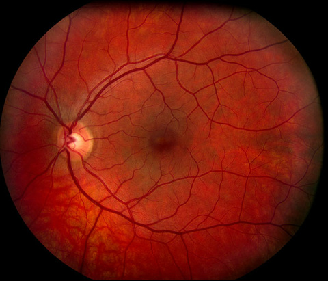
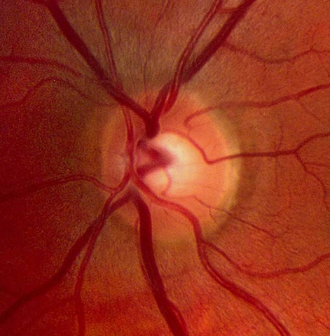
Color Fundus Photography
The Fundus, or inner lining, of the eye is photographed with specially designed cameras through the dilated pupil of the patient. The painless procedure produces a sharp view of the retina, the retinal vasculature, and the optic nerve head (optic disc) from which the retinal vessels enter the eye. The optic disc measures about 1.5mm in diameter.
The vessels form an arc around the macula which produces the central 20 degrees of vision. At the center of the macula lies the tiny fovea, measuring only 500 microns across, which is responsible for our most central reading vision. Color Fundus Photography is used to record the condition of these structures in order to document the presence of disorders and monitor their change over time.
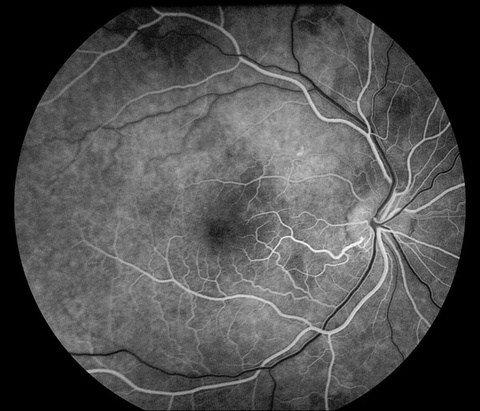
Fluorescein Angiography
Fluorescein angiography was first successfully used in the human eye in 1961* and has evolved since then as one of the fundamental imaging techniques in the eye. It is a test that helps in the differentiation of retinal disease and is used to determine if laser treatment of the retina is warranted.
A contrast medium called Sodium Fluorescein is injected into a vein in the arm. The dye travels quickly through the body's circulatory system, and is photographed in black and white as it travels through the eye. The same camera used for fundus photography is employed for this procedure. Two special filters are used to limit the image to the color of light being emitted from the fluorescent dye.
About twelve seconds after the injection, the dye appears in the arteries of the retina. Over a two to five second period, the dye travels through the very small vessels, or capillaries, and fills the veins. Ten minutes after the injection, the dye has mostly evacuated from the eye, having stained the optic nerve head.
Figures 1-4: Fluorescein Angiogram, Normal
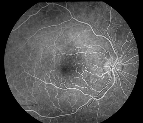
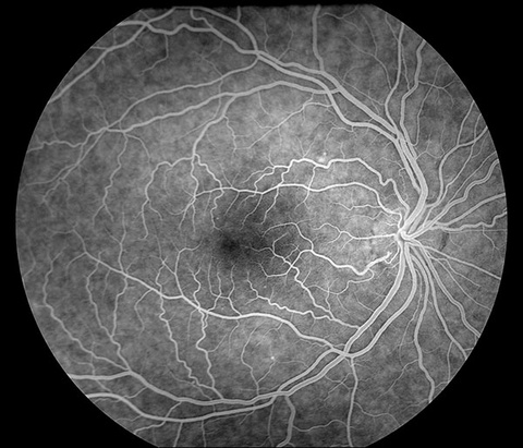

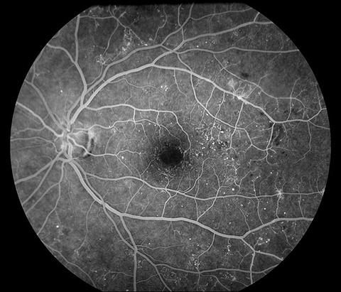
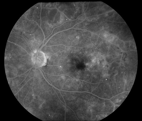
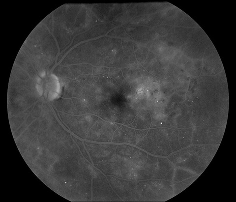
This normal progression of dye is interrupted by many diseases of the choroid, retina, and retinal vasculature. A fluorescein angiogram of a patient with ocular complications due to diabetes (diabetic retinopathy) reveals vascular irregularities when compared with the normal angiogram.
Figures 5-7: Fluorescein Angiogram, Diabetic Retinopathy
Indocyanine Green Angiography
Indocyanine Green Angiography (ICG) is used to acquire an angiogram of the choroid. The choroid is the layer of blood vessels and connective tissue between the sclera (white of the eye) and retina. It supplies nutrients to the inner parts of the eye.
A procedure similar to fluorescein angiography, but ICG angiography uses Indocyanine Green dye, which fluoresces in the infra-red (non-visible) light. The infra-red wavelengths have the ability to penetrate the retinal layers making the circulation in deeper layers visible when photographed with an infra-red sensitive camera.
ICG is injected intravenously and flows through the body to reach the choroidal and retinal circulation. Due to its nature ICG stays in the retinal and choroidal vessels, this allows the distinct outlines of the vessels of the choroid to be seen and identified. ICG is sometimes used to complement fluorescein angiography (FA). FA is often referred to retinal angiography while ICG angiography is referred to choroidal angiography.
ICG was first used in 1969[1] but was not brought into the practical clinical setting until 1992.[2]
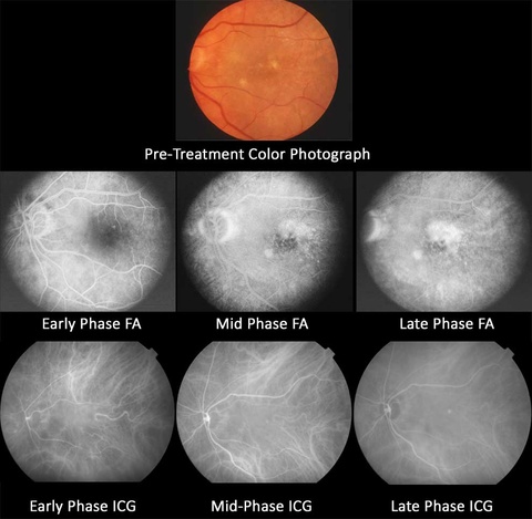
Case Example:
In the case of a patient with 20/100 vision, the fluorescein angiogram demonstrated leakage of fluorescein dye over a large area near the fovea. Traditional treatment would dictate that the entire area of leakage be treated with laser surgery. The treatment of this lesion would cause an instant decline in vision to 20/400.
The ICG angiography, performed on the same day, reveals a pinpoint leak not visible with fluorescein angiography. Focal treatment based on the ICG angiogram caused an increase in vision from 20/100 to 20/80.
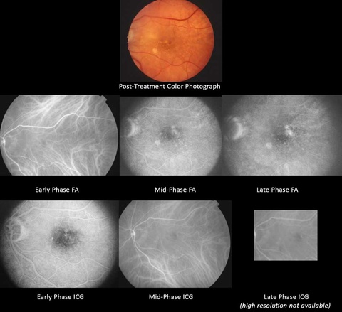
After treatment the laser scar is visible on the color fundus photograph. Both the fluorescein and the ICG angiograms show no leakage of dye demonstrating that the focal treatment was effective.
References:
- Kogure K, Choromokos E. Infrared absorption angiography. J Appl Physiol. 1969;26(1):154-7.
- Yannuzzi LA, Slakter JS, Sorenson JA, Guyer DR, Orlock DA. Digital indocyanine green videoangiography and choroidal neovascularization. Retina.1992;12(3):191-223.
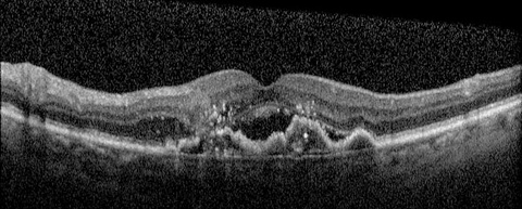
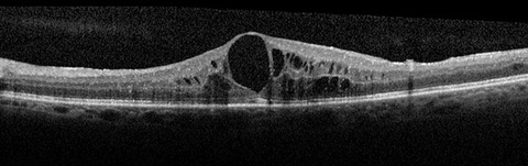
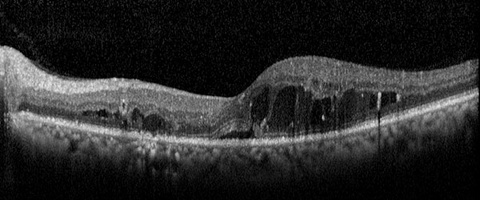
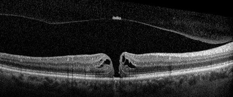
Optical Coherence Tomography (OCT) (Posterior and Anterior)
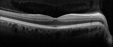
Optical coherence tomography (OCT) is noninvasive, noncontact, imaging technique that can make images of structures in the retina with a resolution of 10 to 17 microns. Cross-sectional images of the retina in a fashion similar to ultrasound but at a much higher resolution and using light waves rather than sound waves.
In OCT, the anatomic layers within the retina can be visualized and the thickness of the retina can be measured. This is an exceptional advancement in diagnostic imaging because it allows real-time visualization of the microstructure of a tissue without the need to excise and process a biopsy specimen. It is used both for diagnosis and to guide intervention.
The OCT is useful in visualizing the pathology of many eye diseases including diabetic retinopathy, macular degeneration, macular holes, epiretinal membranes, macular edema, central serous choroidopathy and optic disc pits.
OCT was first introduced in 1991[Huang, 1991] and has since been adapted to a number of clinical biomedical imaging settings including ophthalmology. Its largest ophthalmology impact has been the imaging of the retina.

Reference: Huang D, et al. Optical coherence tomography. Science. 1991;254(5035):1178-81.
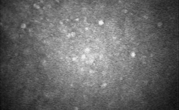
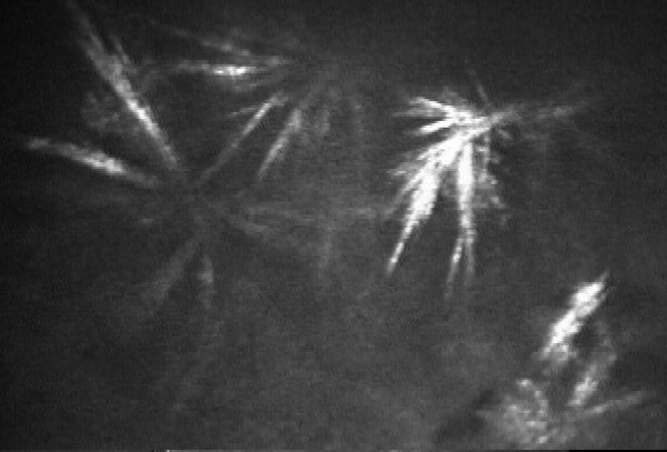
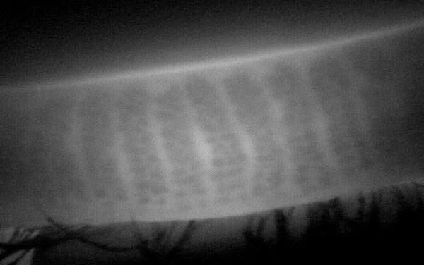
Confocal Microscopy
Tandem Scanning Confocal Microscopy (TSCM) is available through the Corneal Service. TSCM is useful for detecting and managing Keratitis (infections and inflammations of the cornea), especially those caused by acanthamoeba, filamentous fungi, and infectious crystalline keratopathy. TSCM provides a noninvasive method of following resolution of these infections and separating medication side effects from persistent or worsening infections. TSCM is also useful in analyzing corneal dystrophies, tear film abnormalities and nonspecific keratitis.
Corneal and External Photography
Corneal and External Photography in ophthalmology involves capturing detailed images of the eye's external structures, including the cornea, conjunctiva, sclera, and eyelids. This type of photography is crucial for documenting various eye conditions, such as lesions, morphological abnormalities, and nerve anomalies[1].
External photography typically uses conventional cameras to capture images ranging from the entire face to close-ups of the eye. Digital single-lens reflex (DSLR) cameras are commonly used due to their excellent image quality and versatility[1].
Corneal photography often employs specialized equipment like photo slit-lamps to achieve high magnification views of the anterior segment of the eye, including the cornea and iris[2]. These images are essential for diagnosing and monitoring eye diseases and conditions[3].
References
Corneal Topography
Corneal Topography is a non-invasive imaging technique used in ophthalmology to map the surface curvature and shape of the cornea, the clear front window of the eye. This technique works similarly to a 3D map, highlighting features like elevations and depressions on the corneal surface.
Key Uses of Corneal Topography:
- Diagnosing and Monitoring Eye Conditions: It helps detect and track conditions like astigmatism, keratoconus, and corneal scarring.
- Surgical Planning: It is crucial for planning refractive surgeries (e.g., LASIK) and cataract surgeries, ensuring precise reshaping of the cornea.
- Contact Lens Fitting: It aids in fitting contact lenses, especially in cases with significant corneal irregularities.
The procedure is quick and painless, involving the patient looking at a target while images are captured.
References
Operating Room Photography
Operating Room Photography in ophthalmology involves capturing high-quality images during surgical procedures. These images are used for various purposes, including:
- Documentation: Recording the surgical steps and techniques for medical records and future reference[1].
- Education: Providing visual aids for teaching medical students, residents, and other healthcare professionals[2].
- Research: Supporting clinical studies and publications by documenting unique cases and surgical outcomes[3].
Specialized equipment and techniques are used to ensure clear and detailed images, despite the challenging environment of the operating room[3].
References
Slitlamp Biomicroscopy
Slitlamp Biomicroscopy is a fundamental diagnostic technique in ophthalmology that uses a specialized microscope called a slit lamp. This instrument provides a detailed, three-dimensional view of the eye's structures, including the cornea, lens, iris, and anterior chamber[1].
Key Features:
- High Magnification and Illumination: The slit lamp emits a focused beam of light, allowing for precise examination of the eye's anatomy[1].
- Comprehensive Examination: It helps in diagnosing a wide range of eye conditions, such as cataracts, corneal injuries, and retinal issues[2].
- Versatility: With additional lenses, it can also be used to examine the posterior segment of the eye[1].
The procedure is quick and non-invasive, making it an essential tool for routine eye exams and emergency evaluations[2].
References
Ocular evaporation, infrared meibography and tear flow analysis.
Multiple methods of analyzing the ocular surface and tear film for abnormalities are available through the Corneal Service. Ocular evaporation is useful in defining the cause for symptoms of dry eye. Infrared meibography provides detailed analysis of the meibomian gland architecture without the delay of infrared photography. Meibomian glands may be abnormal in occlusive meibomitis, ocular rosacea or seborrhic blepharitis. Tear flow analysis is a specific measure of tear volume and manufacture without the stimulation of test strips (Shirmer's test). All of these methods are used to define the causes of dry eyes and blepharitis to improve both detection and treatment.

Wide Field Fundus Photography and Angiography
Wide Field Fundus Photography and Angiography are advanced imaging techniques used in ophthalmology to capture detailed images of the retina, the light-sensitive tissue at the back of the eye.
Wide Field Fundus Photography:
- Captures Extensive Retinal Views: This technique provides a broader view of the retina compared to traditional fundus photography, allowing for better detection and monitoring of peripheral retinal diseases[1].
- Non-Invasive: It involves taking high-resolution images of the retina using a specialized camera, which helps in diagnosing conditions like diabetic retinopathy, retinal detachment, and age-related macular degeneration[2].
Wide Field Angiography:
- Enhanced Visualization of Blood Vessels: This technique involves injecting a fluorescent dye into the bloodstream and capturing images as the dye travels through the retinal blood vessels. It helps in identifying abnormalities in blood flow and vascular structures[1].
- Critical for Diagnosing Vascular Diseases: It is particularly useful for diagnosing and managing conditions like retinal vein occlusion, diabetic retinopathy, and choroidal neovascularization[1].
These techniques are essential for comprehensive retinal evaluation and play a crucial role in the early detection and management of various eye diseases[1].
References
Contact Information
Ophthalmic Diagnostic Imaging 319-356-0392
Ophth-Photographers@healthcare.uiowa.edu
For information about different types of Ophthalmic Photography and a more in-depth description of methods and principles, see The Ophthalmic Photographer's Society website.